Forehead Osteoma Removal
Forehead osteoma removal is a procedure where the cosmetic result depends almost entirely on where the incision is placed and how it’s closed — removing the bony growth itself is the simpler part of the operation. Osteomas are benign, slow-growing bony masses that most commonly form as hard knobs on the forehead, particularly in the area between the hairline and the eyebrow. They are typically asymptomatic — though some patients report facial pain or headaches — and the most frequent concern is their unsightly visible appearance. Forehead osteomas may be present at birth or develop slowly over many years. While they rarely cause functional problems, they can significantly affect the overall aesthetics and symmetry of the face. Forehead osteoma removal is one of the procedures within facial reconstruction and aesthetics at Dr. Khosh’s Park Avenue practice in New York City.
What separates a satisfying forehead osteoma removal from a noticeable one is the surgeon’s command of endoscopic technique and hairline-hidden incision placement. Dr. Maurice Khosh has spent three decades performing endoscopic procedures across the forehead, brow, and midface region. Dual board-certified by the American Board of Facial Plastic and Reconstructive Surgery and the American Board of Otolaryngology–Head and Neck Surgery and a Fellow of the American College of Surgeons (FACS), he was the youngest recipient of Columbia University Medical Center’s Maxwell Abramson Resident Teaching Award. Dr. Khosh has been recognized as a perennial Castle Connolly Top Doctor.
The Endoscopic Removal Technique
Forehead osteoma removal usually involves a classic minimally invasive approach: a small buttonhole incision is made at the hairline or hidden within the scalp, and an endoscope is used to visualize the bony growth and the surrounding anatomy. The osteoma is then carefully contoured down to the level of the surrounding bone using specialized instrumentation. The procedure does not usually require deep perforation of the skull — the majority of forehead osteomas involve only the outer portion of the cranial bone (the outer table), allowing the surgery to be performed safely from a superficial plane. The endoscopic visualization combined with the carefully positioned incision allows for swift removal — typically less than 30 minutes — with minimal recovery time and no visible scarring once the hair grows in over the closure.
Case Study
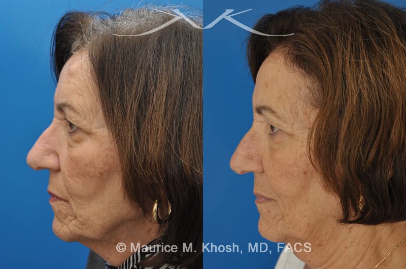
This 65 year old who had noted a progressively enlarging hard mass in the left upper forehead. The clinical examination finding was consistent with an osteoma. The osteoma was successfully removed as an office procedure utilizing local anesthesia. The incision was perfectly hidden inside the hairline above the location of the osteom. The incision was designed to allow hair growth through the scar, thereby camouflaging it.
- Patient: 68 year old lady interested in osteoma removal in Manhattan
- Problem: Hard, round, raised mass underneath the forehead skin
- Procedure: Osteoma removal as an office procedure, under local anesthesia
Disclaimer: These are actual results for patients of Dr. Maurice Khosh. Plastic and cosmetic surgery results can vary between patients.
What to Expect from the Procedure
Forehead osteoma removal is typically performed in Dr. Khosh’s Park Avenue office under local anesthesia, with the procedure taking less than thirty minutes from start to finish. After cleansing and numbing the area, Dr. Khosh makes a small buttonhole incision hidden within the hairline above the osteoma. Using endoscopic visualization, he carefully contours the bony growth back to the natural surrounding contour. The incision is closed with fine sutures designed to allow hair growth through the closure line, gradually camouflaging the scar entirely within the hairline. Patients are sent home the same day and most resume normal activities immediately, with mild swelling and minor bruising typically resolving within the first one to two weeks.
“The art of forehead osteoma removal is not the bone work — it’s the incision placement and the closure technique. Done well, the result is a smooth, symmetric forehead with no visible evidence that any surgery was performed. The endoscopic approach is what makes that possible, but only when the incision is positioned to allow the hair to fully camouflage the closure.” — Dr. Maurice Khosh
Why a Small Incision Matters
The most important variable in the cosmetic outcome of forehead osteoma removal is the size and position of the incision. A larger, traditional incision can leave a visible scar regardless of how well the osteoma is removed. The buttonhole endoscopic approach uses an incision small enough to be entirely hidden within the hair, with the additional advantage that the design allows the hair follicles to continue growing through the healed scar — making the closure invisible to anyone, including the patient. This is the key advantage of the endoscopic technique over older surgical approaches.
Why Patients Choose Dr. Khosh for Forehead Osteoma Removal
- Endoscopic Surgical Expertise: Three decades of endoscopic procedures across the forehead and midface region
- Hidden Incision Technique: Hairline incision designed to allow hair growth through the closure, leaving no visible scarring
- Office-Based Procedure: Most osteoma removals performed under local anesthesia with same-day return to normal activities
- Maxwell Abramson Resident Teaching Award: Youngest recipient at Columbia University Medical Center
- Dual Board Certification: Combined facial plastic and head and neck surgery expertise
- Park Avenue Convenience: Private Upper East Side practice serving patients from across Manhattan and the tri-state area
Schedule Your Consultation in Manhattan
If you are living with a forehead osteoma and would like to restore the natural symmetry and contour of your forehead, the endoscopic removal procedure can produce a scar-free result in a short office-based visit. To schedule a private consultation with Dr. Khosh at his Park Avenue office in New York City, contact our office today, or call (212) 339-9988.
Minimally Invasive Osteoma Removal Patient Testimonial

Click Here to See Osteoma Removal Photo Gallery